X-Ray VS MRI: Which is Better?
X-rays and MRI are complementary studies.

Both are useful in different ways.
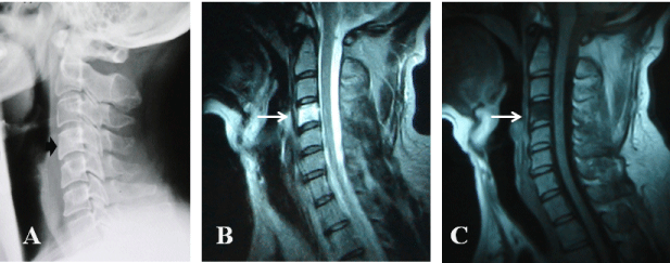
X-rays are fairly simple to do and are excellent in showing the bones. They are also good in showing degenerative changes, like disk arthritis and fractures. X-rays are helpful for showing alignment and any instability of the spine on flexion and extension views, especially in the lumbar spine. They are not so good at showing soft tissue injuries, like disk herniations, although in the neck you may see soft tissue swelling that can alert the physician to a subtle neck fracture.
An MRI, on the other hand, is better at showing soft tissue injuries, such as disk herniations. They are not as good at showing gross instability in the spine that you would see on flexion extension X-rays. There are flexion-extension MRI that can show gross instability or abnormal movement of the vertebral bodies, but they are not common and usually unnecessary if X-rays are done. Also, an MRI can be confusing when looking at an arthritic spine because a soft tissue disk herniation and a bony spur can look similar. A complementary X-ray can help clear up that confusion.
With fractures of the spine, both are helpful in showing the extent of the injury. Surgical indications are usually made on X-ray but the MRI can show other soft tissue damage that is very helpful when deciding on best surgical approach.
In short, both x-rays and MRI are useful when diagnosing a patient because they look at the spine in different but complementary ways. It is a bit like a CSI crime scene where regular photos are important but a black light can show other important factors that photos don’t show. Both are needed to figure it out. You start with the simple thing that gives a lot of information like a photo (or X-ray) and add on other studies like an MRI when you decide you need more information.
